Home | About | Faculty | Calendar | Facilities | Graduate program | Contact | Apply
This page is optimized for viewing with javascript.
Bruce A. Bowerman

Professor, Biology
Member, IMB
Ph.D., University of California, San Francisco
B.A., Kansas State University
Email
Lab website
Office: Lewis Integrated Science Building 314
Office Phone: 541-346-0853
Lab: Lewis Integrated Science Building 307
Lab Phone: 541-346-4551
Loading profile for Bruce A. Bowerman
Research Interests
Worm Breeder’s Gazette Profile
The Bowerman lab uses molecular genetics and live cell imaging to study cytoskeletal regulation and function in the early Caenorhabditis elegans embryo. Beginning with the first mitotic division, the early embryo undergoes a sequence of five asymmetric cleavages. These early divisions are largely responsible for establishing the pattern of cell fates required for normal early embryonic development. These asymmetric cell divisions, with their highly stereotyped timing and mitotic spindle positioning, provide a rich context in which to use the powerful genetics of C. elegans to investigate cytoskeleton function during cell division and development.

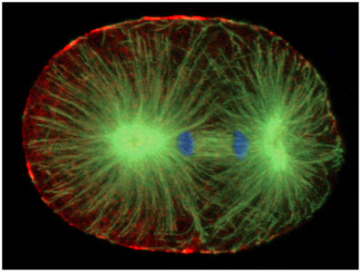
The actomyosin cytoskeleton, including the non-muscle myosin II called NMY-2 (in red in the late anaphase mitotic one-cell stage embryo shown above) is localized predominantly to the cell cortex. The actomyosin cytoskeleton is important both for generating anterior-posterior polarity, and for cytokinesis. Microtubules (in green in the above figure; DNA is in blue) form both the meiotic and mitotic spindles, which capture and segregate chromosomes during cell division.
Current projects in lab focus on both oocyte meiotic spindle assembly, which occurs in the absence of the microtubule organizing centers called centrosomes, and mitotic spindle assembly, which is organized in large part by centrosomes. The movie below on the left shows Meiosis I and II in an oocyte after fertilization and during ovulation. A row of three oocytes are present on the left, with the spermathecum adjacent to the most mature oocyte, and mitotic embryos in the uterus are visible at the right. This movie was filmed using whole mount worms expressing a GFP fusion to tubulin to label microtubules in green, and an mCherry fusion to a histone to label chromosomes in red. The movie below on the right shows the first two rounds of mitotic division in an isolated early embryo (again with GFP labeling microtubules in green, and mCherry labeling histones in red).
Recent publications
(pulled from pubmed)
Recent publications
(pulled from pubmed)
Du Z, He F, Yu Z, Bowerman B, Bao Z
Dev Biol 2015 Feb 15;398(2):267-79
Phillips PC, Bowerman B
Curr Biol 2015 Mar 16;25(6):R223-5
Lowry J, Yochem J, Chuang CH, Sugioka K, Connolly AA, Bowerman B
G3 (Bethesda) 2015 Aug 26;
Connolly AA, Sugioka K, Chuang CH, Lowry JB, Bowerman B
J Cell Biol 2015 Sep 14;210(6):917-32
Connolly AA, Osterberg V, Christensen S, Price M, Lu C, Chicas-Cruz K, Lockery S, Mains PE, Bowerman B
Mol Biol Cell 2014 Apr;25(8):1298-311
Keikhaee MR, Nash EB, O'Rourke SM, Bowerman B
PLoS One 2014;9(9):e106484
De Arras L, Seng A, Lackford B, Keikhaee MR, Bowerman B, Freedman JH, Schwartz DA, Alper S
J Biol Chem 2013 Jan 18;288(3):1967-78
Burger J, Merlet J, Tavernier N, Richaudeau B, Arnold A, Ciosk R, Bowerman B, Pintard L
PLoS Genet 2013 Mar;9(3):e1003375
Schneider SQ, Bowerman B
Curr Biol 2013 Apr 22;23(8):R313-5
Bowerman B, O'Rourke SM
Science 2012 May 25;336(6084):984-5
O'Rourke SM, Carter C, Carter L, Christensen SN, Jones MP, Nash B, Price MH, Turnbull DW, Garner AR, Hamill DR, Osterberg VR, Lyczak R, Madison EE, Nguyen MH, Sandberg NA, Sedghi N, Willis JH, Yochem J, Johnson EA, Bowerman B
PLoS One 2011 Mar 1;6(3):e16644
O'Rourke SM, Yochem J, Connolly AA, Price MH, Carter L, Lowry JB, Turnbull DW, Kamps-Hughes N, Stiffler N, Miller MR, Johnson EA, Bowerman B
Genetics 2011 Nov;189(3):767-78
Bowerman B
Mol Biol Cell 2011 Oct;22(19):3556-8
O'Rourke SM, Christensen SN, Bowerman B
Nat Cell Biol 2010 Dec;12(12):1235-41
Dorfman M, Gomes JE, O'Rourke S, Bowerman B
Genetics 2009 Aug;182(4):1035-49
Gillis WQ, St John J, Bowerman B, Schneider SQ
BMC Evol Biol 2009 Aug 20;9:207
Gillis WQ, Bowerman BA, Schneider SQ
BMC Evol Biol 2008 Apr 15;8:112
Canman JC, Lewellyn L, Laband K, Smerdon SJ, Desai A, Bowerman B, Oegema K
Science 2008 Dec 5;322(5907):1543-6
Gillis WJ, Bowerman B, Schneider SQ
Evol Dev 2007 Jan-Feb;9(1):39-50
Schneider SQ, Bowerman B
Dev Cell 2007 Jul;13(1):73-86
O'Rourke SM, Dorfman MD, Carter JC, Bowerman B
PLoS Genet 2007 Aug;3(8):e128
Goulding MB, Canman JC, Senning EN, Marcus AH, Bowerman B
J Cell Biol 2007 Sep 24;178(7):1177-91
Willis JH, Munro E, Lyczak R, Bowerman B
Mol Biol Cell 2006 Mar;17(3):1051-64
Bowerman B, Kurz T
Development 2006 Mar;133(5):773-84
Bowerman B
Curr Biol 2006 Dec 19;16(24):R1039-42
Bowerman B
Science 2005 May 20;308(5725):1119-20
Bowerman B
Cell 2005 Jun 3;121(5):662-4